Fast Volumetric Imaging of Seizures with Adaptive Optics Light Sheet Microscopy
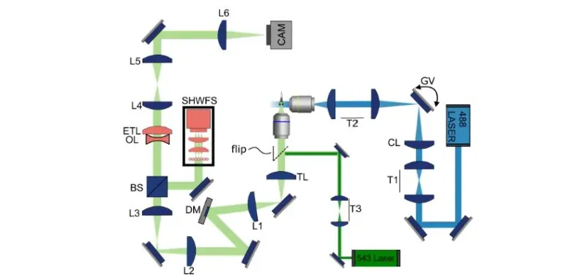
Generally this is done using light sheet microscopy, which provides effectively a 2D ‘slice’ of the tissue in question, but to observe a brief event in a larger biological system you need to be able to rapidly change the layer and focus between the virtual layers. This is what [Bingxi Liu] et al. al did using adaptive optics with an electrically tunable lens (ETL) in order to capture seizures in the brain of zebrafishes. Their system can capture a volume of 499 × 499 × 150 μm3 at 4 volumes per second, which is large enough to fit optically transparent zebrafish larva into.
- ▪Generally this is done using light sheet microscopy, which provides effectively a 2D ‘slice’ of the tissue in question, but to observe a brief event in a larger biological system you need to be able to rapidly change the layer and focus bet
- ▪This is what [Bingxi Liu] et al. al did using adaptive optics with an electrically tunable lens (ETL) in order to capture seizures in the brain of zebrafishes.
- ▪Their system can capture a volume of 499 × 499 × 150 μm3 at 4 volumes per second, which is large enough to fit optically transparent zebrafish larva into.
Hackaday files mainly under tech. We currently carry 319 of its stories.
Story provenance
Source · retrieval · rights · ranking — open for full record
inspect →
Story provenance
Attribution is not the same as permission. This drawer separates discovery metadata, excerpts, WeSearch-generated summaries, reuse status, and whether the publisher receives the visit. Nothing here claims a legal grant the publisher has not made.
Record
| Original publisher | Hackaday |
| Canonical URL | https://hackaday.com/2026/07/24/fast-volumetric-imaging-of-seizures-with-adaptive-optics-light-sheet-microscopy/ |
| Publication time | Sat, 25 Jul 2026 02:00:08 +0000 |
| Retrieval time | 2026-07-25T02:05:42.225Z |
| Last seen | 2026-07-25T02:05:42.225Z |
| Headline source | Publisher (no WeSearch rewrite) |
| Excerpt source | publisher body |
| Excerpt method | First ~120 words (~800 chars) of extracted publisher body, fair-use limited. |
| Summary | WeSearch · cerebras-chat (WeSearch summarizer) |
| Summary source text | contentText |
| Citation coverage | Summary is a WeSearch-generated derivative; primary citation is the original publisher URL. |
| Cluster | 7PsZVpVKGHYE |
| Cluster logic | Grouped by semantic title/content similarity across sources within a rolling window. Same-publisher template collisions are excluded from coverage comparison. |
| Ranking reason | Story pages are not engagement-ranked. Hub feeds use recency, with optional source-diversified chronological ordering (cap consecutive stories per source). No personalized ranking. |
| Publisher visit | Yes — open original |
| Substitutes article? | No — link-out required for full text |
Rights status (four layers)
WeSearch handling by dimension
| Indexing | May the item be indexed (stored, ranked, made findable)? | Allowed |
| Snippet | May a short excerpt of the publisher's text be shown? | Allowed |
| AI summary | May WeSearch generate its own short summary of the article? | Limited |
| Retrieval / RAG | May the content be exposed for third-party retrieval-augmented generation? | Not asserted |
| Model training | May the content be used to train AI models? | Not asserted |
| Commercial reuse | May the content be reused commercially? | Not permitted |
Basis: Derived from the published RSS/Atom feed. Contact: [email protected]. Reviewed: 2026-07-24.
Opening excerpt (first ~120 words) tap to expand
Fast Volumetric Imaging Of Seizures With Adaptive Optics Light Sheet Microscopy No comments by: Maya Posch July 24, 2026 Title: Copy Short Link: Copy Seizure in zebrafish larva imaged using AO setup. (Credit: Bingxi Liu et al., Biomedical Optics Express, 2026) Key to understanding something like epilepsy is to be able to record highly transient events in biological tissues. Generally this is done using light sheet microscopy, which provides effectively a 2D ‘slice’ of the tissue in question, but to observe a brief event in a larger biological system you need to be able to rapidly change the layer and focus between the virtual layers. This is what [Bingxi Liu] et al.
…
Excerpt limited to ~120 words for fair-use compliance. The full article is at Hackaday.
Discussion
0 commentsMore from Hackaday